


Special Cases
Special cases that are instructional and illustrative
of typical zoo dentistry cases
Siberian tiger mandibular canine endodontic procedure
October 16, 2014
Milwaukee County Zoo.
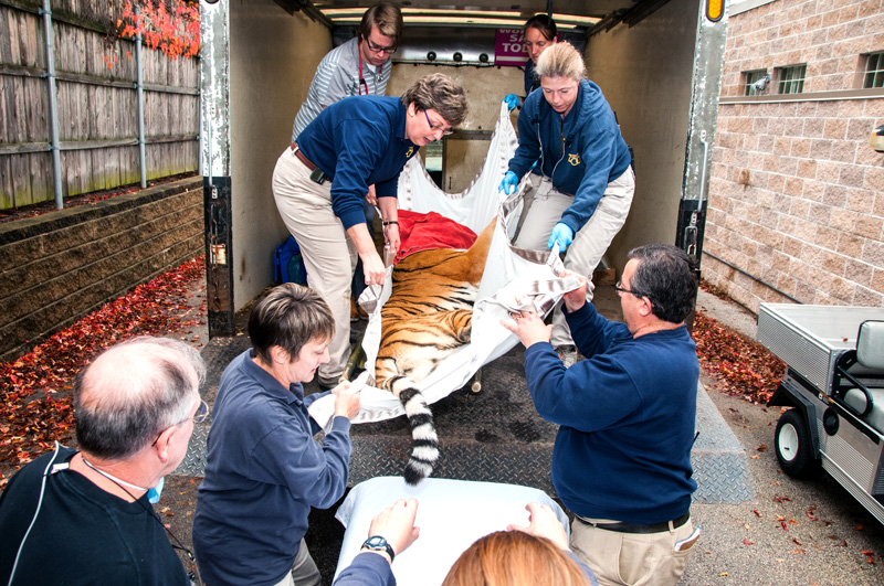
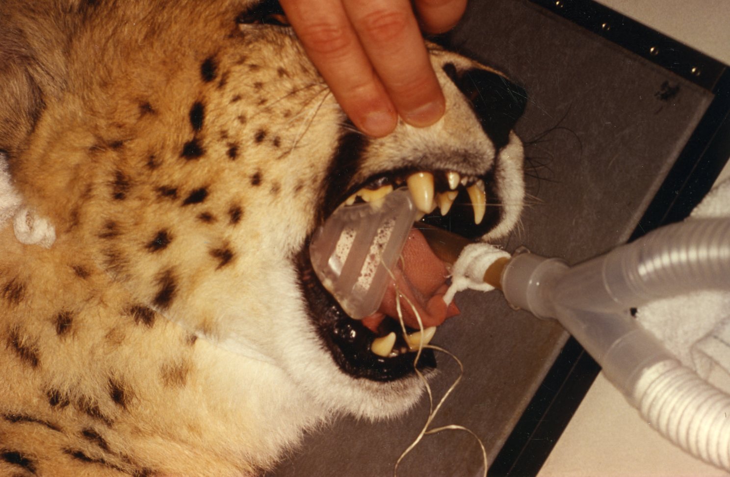
Transporting Tula
(Click image to enlarge)
photo by Mike Nepper
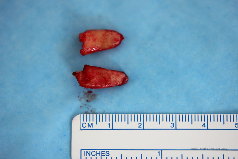
"Tula" is a 2 1/2 year old Siberian tiger, weighing 242 pounds. She fractured her mandibular canine when playing with her littermate, sister, on an elevated structure. An observant zookeeper spotted the canine tip piece, approximately 1 1/2 cm. long, shortly after the incident and confirmed by observing the tigress, that the canine was short, fractured.
Tula did not seem to be in pain or distress and her behavior was normal according zookeepers. However, I am aware of fresh fractures of this type, exposing the pulp and nerve caused some captive carnivores to be in so much pain and distress that they attacked and killed a cage mate. ("Bubba", Brookfield zoo, year?)
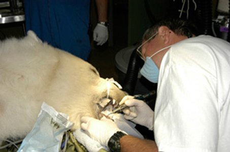
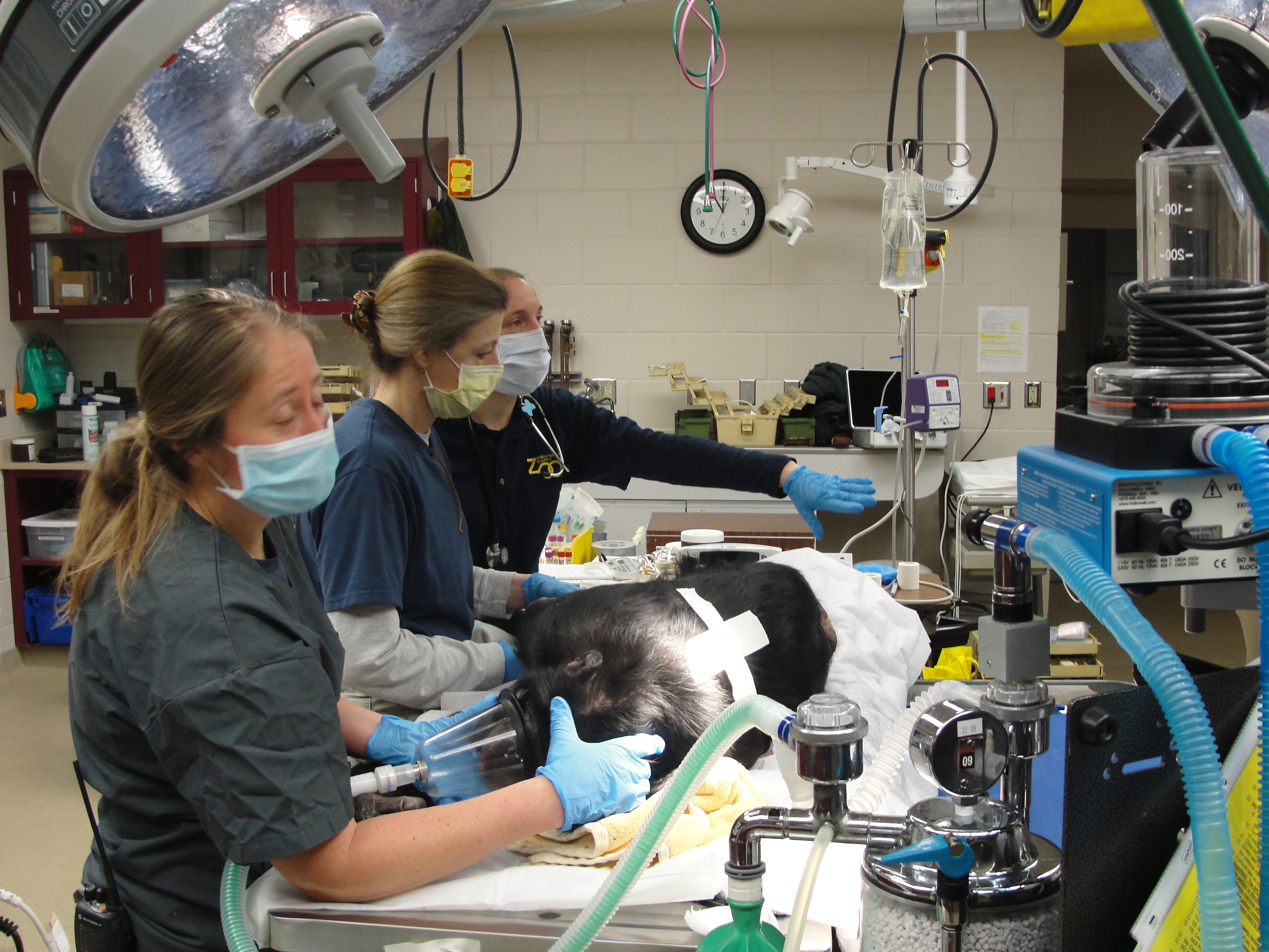
Approximately ten days later, on October 16, 2014 the zoo veterinarians, primarily Drs. Jennifer Haussmann and Vickie Clyde, sedated Tula, and transported her to the Milwaukee County Zoo Animal Health Center. Following the vets and techs establishing an intravenous line, attaching a pulse-oximeter and intubation establishing general anesthesia, we were given permission to start the dental procedure.
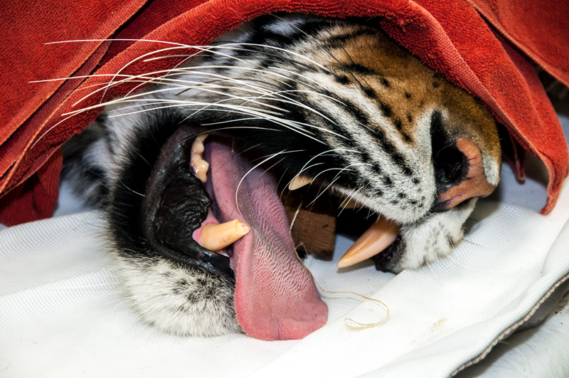
Figure 1
(Click image to enlarge)
photo by Mike Nepper
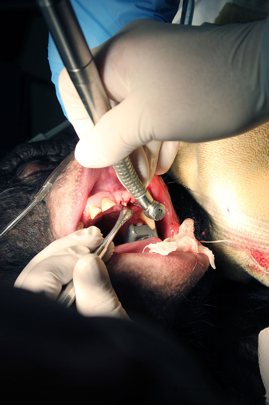
A brief oral exam revealed no other oral or dental pathology warranting treatment other than the mandibular right canine. The pulp was exposed with pulp tissue at the surface of the opening. The fractured canine segment and observation of the tooth had already established the pulp exposure (Figure 1).
The pulp was vital. The preoperative radiograph (Figure 2) revealed the pulp on this young animal widened a short distance beyond the fracture.
After explaining to the veterinarians, I used the high speed dental hand piece to cut another approximate cm off of the tooth to permit optimal pulp access for the endodontic procedure (Figures 3 and 4).
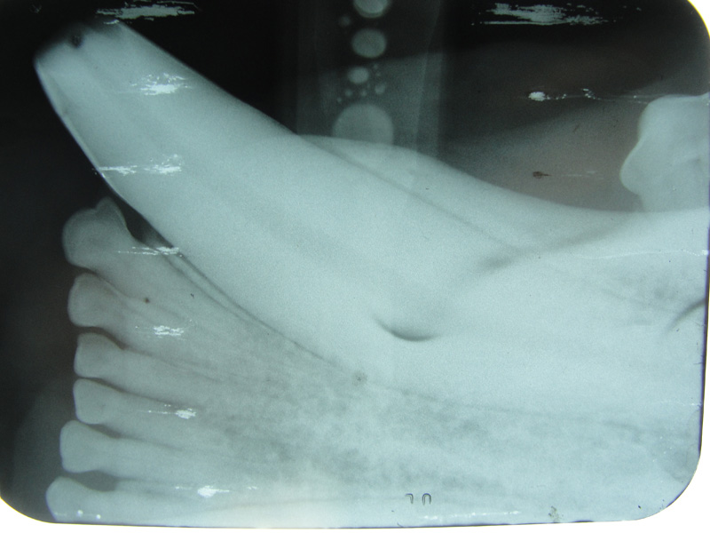
Figure 2
(Click image to enlarge)
photo by Mike Nepper
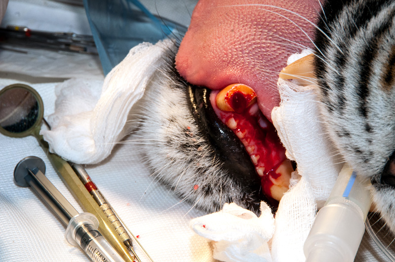
Figure 3
(Click image to enlarge)
photo by Mike Nepper
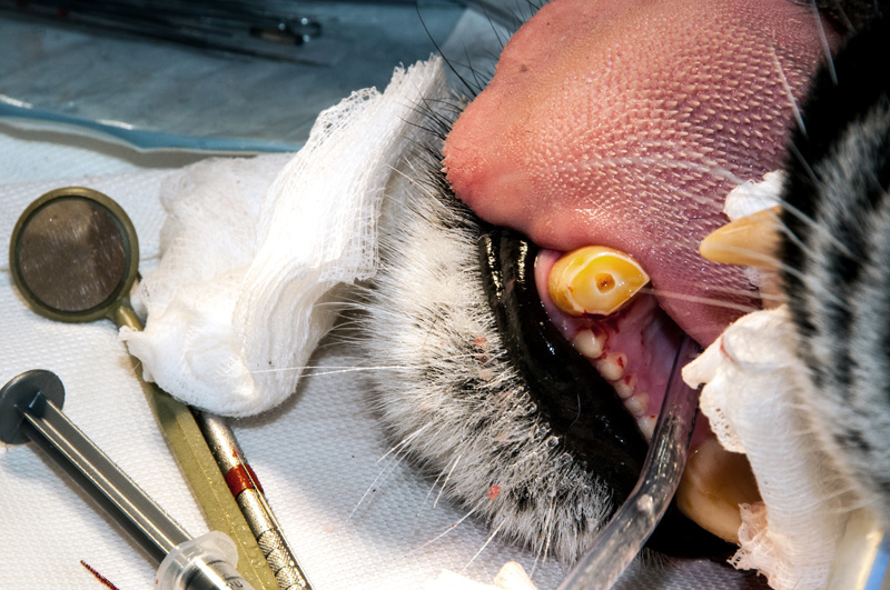
Figure 4
(Click image to enlarge)
photo by Mike Nepper
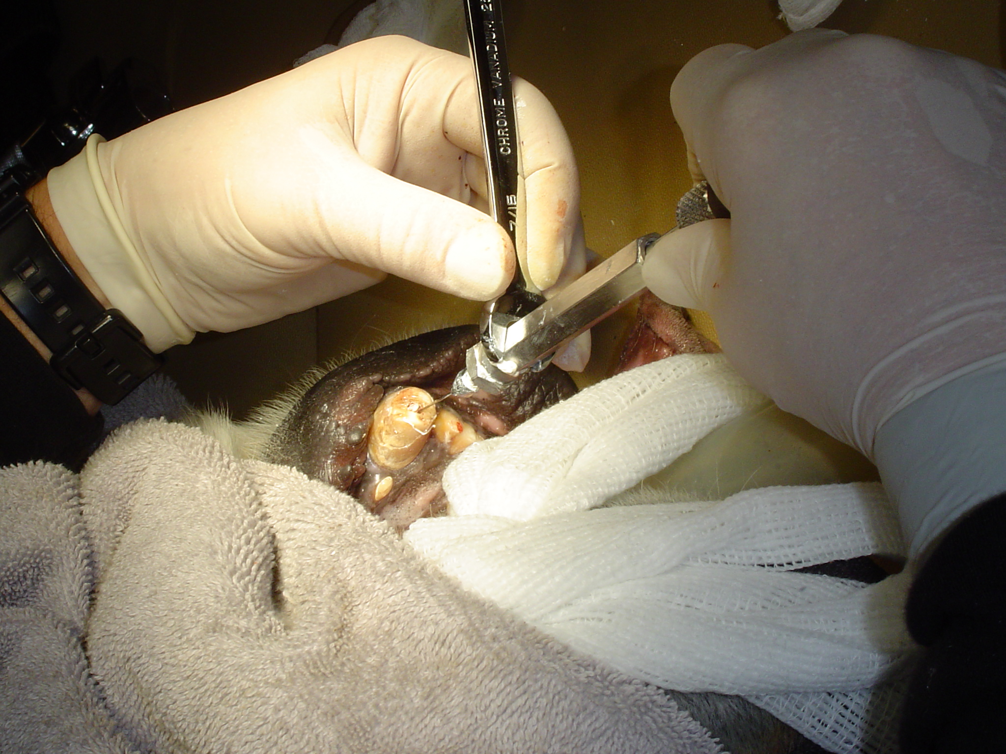
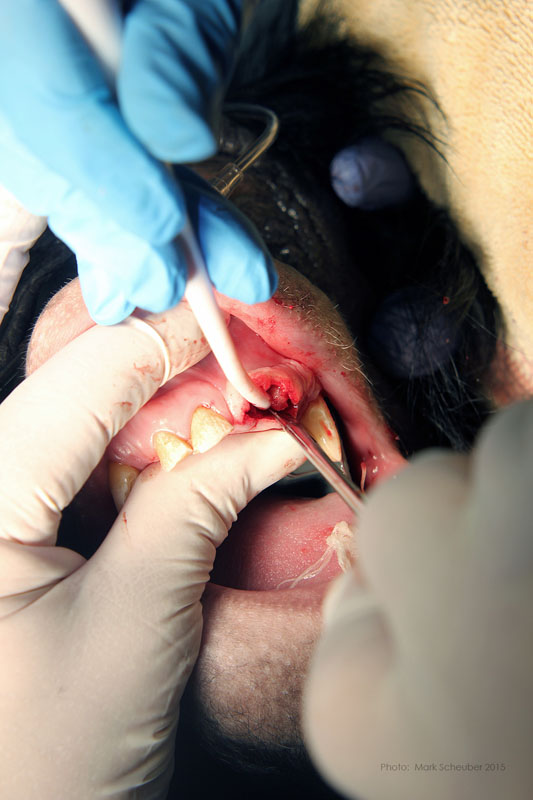
I extirpated the pulp and nervous tissue with a 100 mm endo file. Working length was established at 72 mm. This correlated with my measurement and estimation from the preoperative radiograph. Using successively larger diameter files, and copious irrigation with NaOCl and RC prep the canal was shaped and dissenfected (Figures 5, 6 and 7).
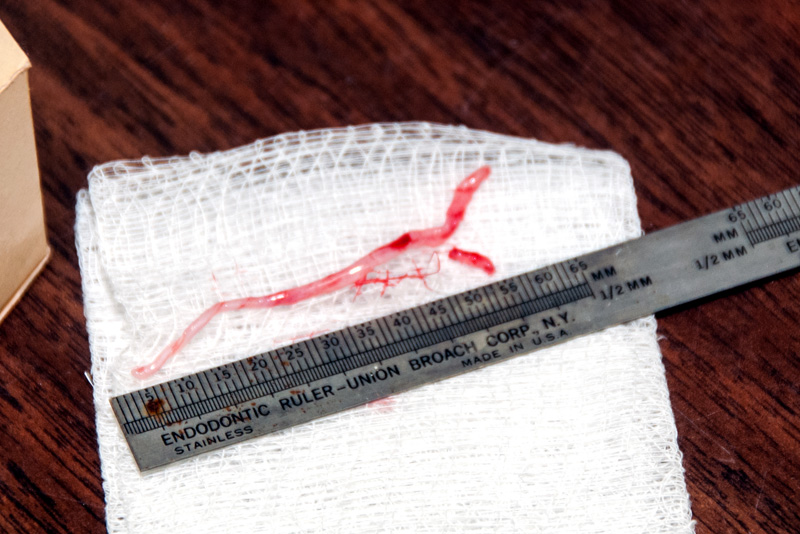
Figure 5
(Click image to enlarge)
photo by Mike Nepper
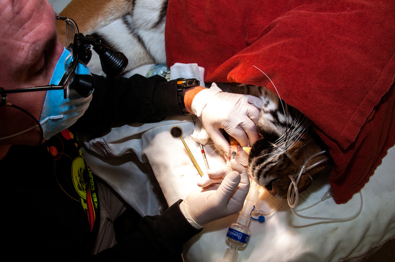
Figure 6
(Click image to enlarge)
photo by Mike Nepper
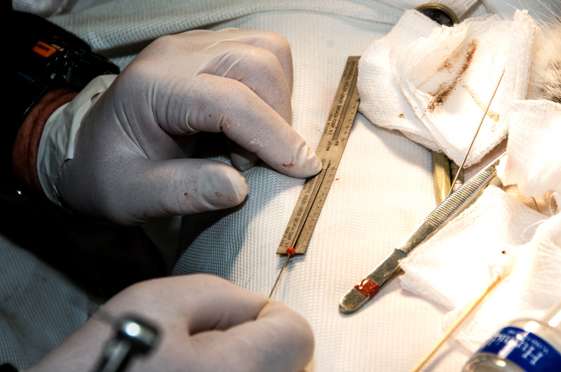
Figure 7
(Click image to enlarge)
photo by Mike Nepper
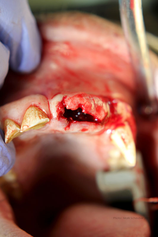
Hemostasis was established and the pulp was dried with sterile pipe cleaners. They must be actual pipe cleaners comprised of absorbent cotton. Not colorful craft items. Bag and sterilize (Figure 8).
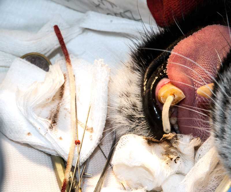
Figure 8
(Click image to enlarge)
photo by Mike Nepper
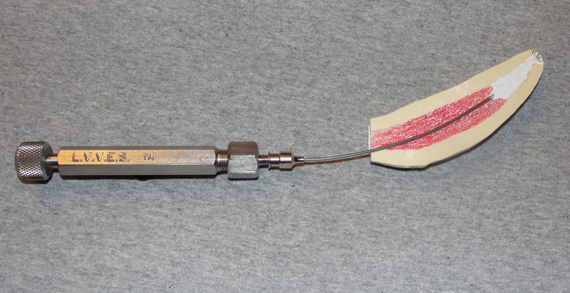
After fitting the master gutta percha cone at the established working length, the canal was then obturated with PCA endo filler paste (a ZOE based paste) by placing the Large Volume Veterinary Endodontic Syringe needle at the apex and backfilling as it is withdrawn. The paste was deposited with a 20 gauge, 2 1/2 inch spinal needle (Figure 9). Additional gutta percha points were then placed and condensed in the canal.
A zinc phosphate cement base was placed over the endo fill. The access opening was briefly prepared and a composite resin restoration placed. I rounded and smoothed off the fractured canine edges when polishing the composite resin (Figure 10).
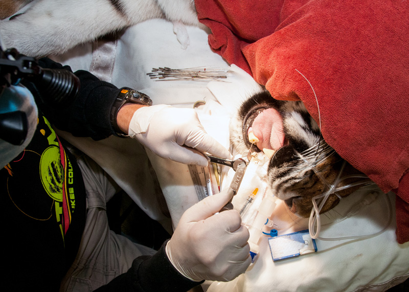
Figure 9
(Click image to enlarge)
photo by Mike Nepper
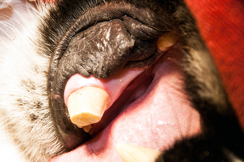
Figure 10
(Click image to enlarge)
photo by Mike Nepper
The entire dental procedure was completed in 53 minutes. Tula recovered from her anesthesia uneventfully. A successful procedure facillitared by excellent general anesthesia by the veterinarians. See the fill radiograph (Figure 11).
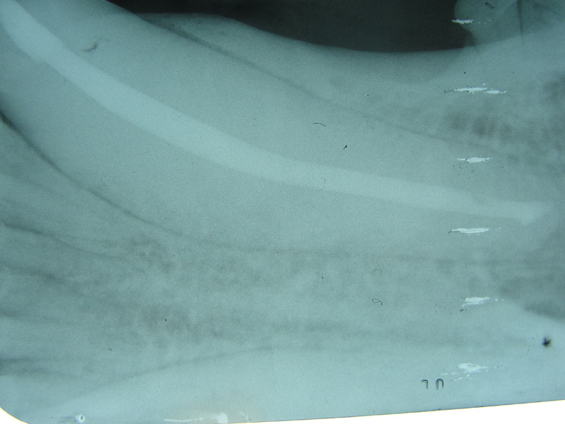
Figure 11
(Click image to enlarge)
photo by Mike Nepper
This conventional endo approach also called coronal or orthograde is certainly the technique of choice for a fresh, vital pulp, endodontic procedure. Considering the delta apex on mature carnivore teeth, if this tooth was abscessed and or a significant amount of lysis of the apex or a large lytic area in the adjacent alveolar bone was observed, a surgical retrograde approach would have been necessary to treat properly. Also, again considering the delta apex of the tooth, periodic examination including radiography of the tooth root is necessary to monitor the success of the conventional endodontic procedure. The necessity of such follow up should be indicated in the patient's chart so that if she must be sedated for some other reason, the dental exam and radiographs will be done also.
Zookeepers are reminded to be particularly observant of behavior or signs that may indicate pain or abscessation of the tooth.
Bonobo Makanza
February, 2015
Milwaukee County Zoo.
Photos by Mark Scheuber, Zoo Keeper/Photographer
(Click image to enlarge)
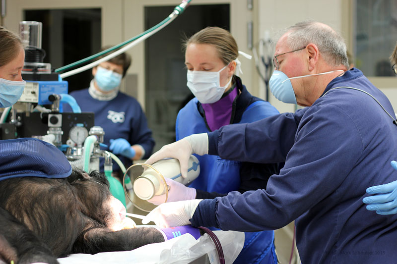
Bonobo Makanza, sedated, being prepared for dental treatment. L to R, zookeeper Stacy Whitaker, hospital keeper Celi Jeske and Veterinarian Dr. Jennifer Hausmann.
(Click image to enlarge)
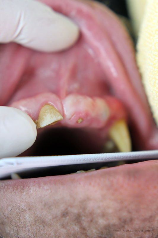
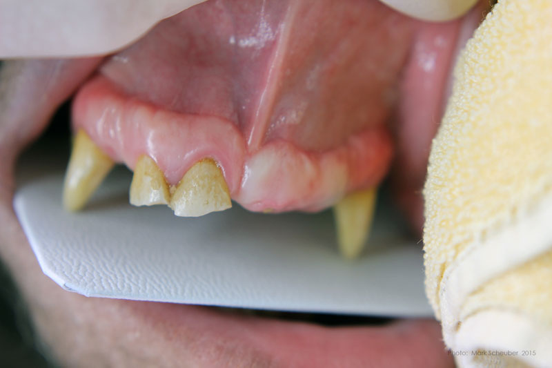
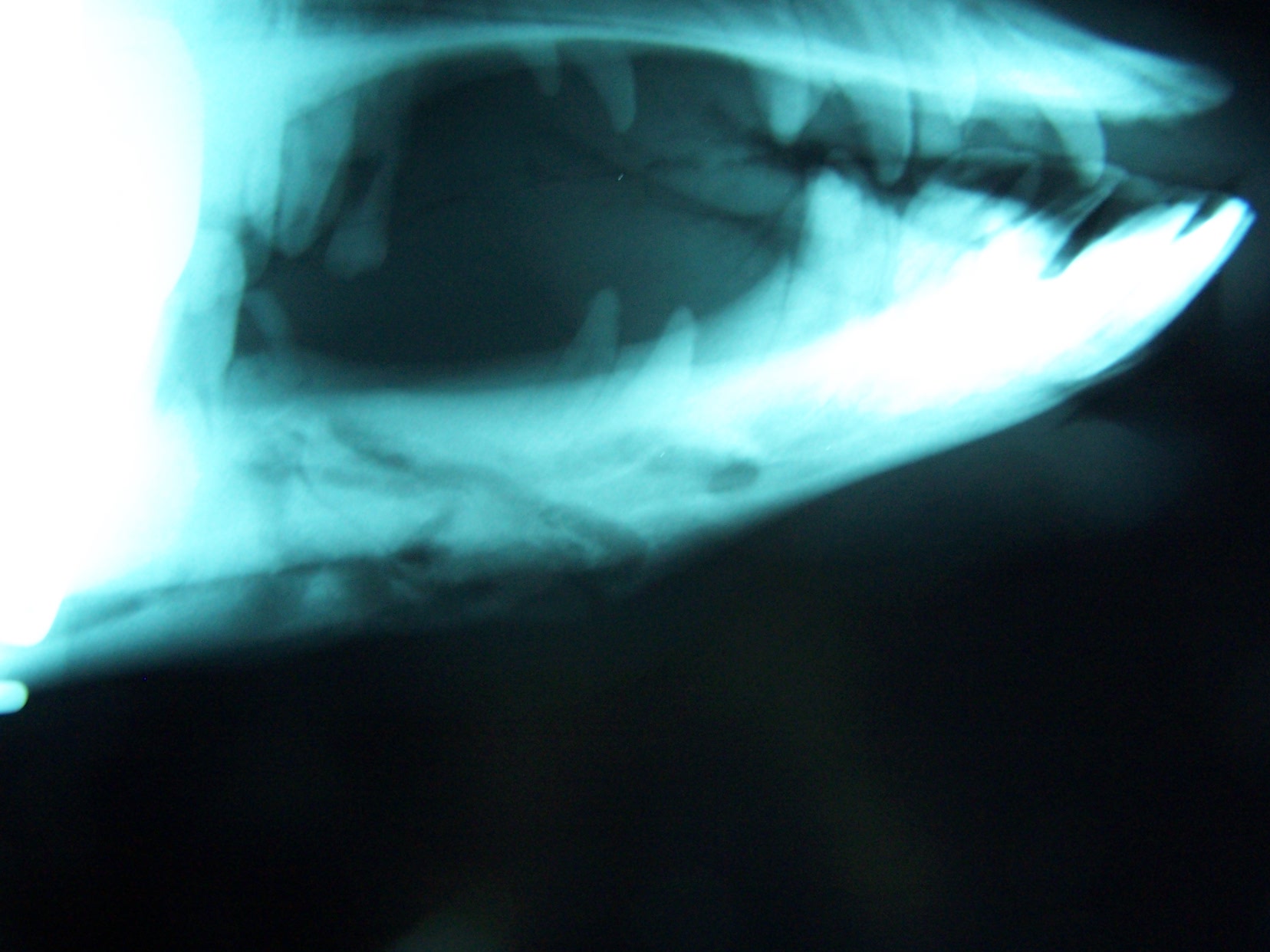
Maxillary left central incisor and lateral incisor residual roots. Region was inflamed, edematous prior to two weeks of antibiotic therapy.
(Click image to enlarge)
Conventional dental film in position. Digital Phos Phor plates were also exposed.
(Click image to enlarge)
Dr. Hausmann and Dr. Scheels positioning Nomad radiography unit.
(Click image to enlarge)
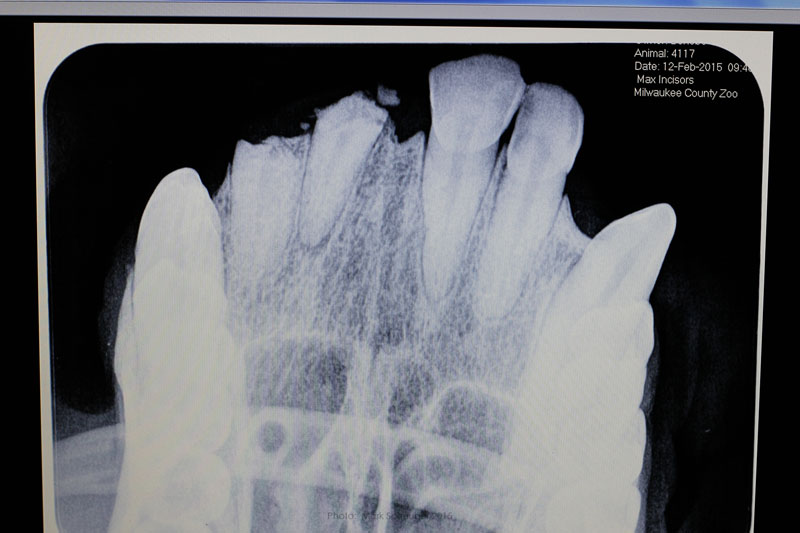
Digital image reveals periapical lesion on central incisor residual root.
(Click image to enlarge)
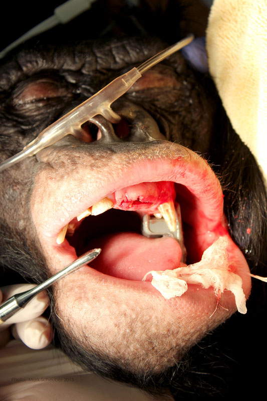
Incision on crest of ridge.
(Click image to enlarge)
556 surgical bur used to remove approximately 2mm. of alveolar bone surrounding each root ("ditching".)
(Click image to enlarge)
Luxator technique utilized to remove residual roots.
(Click image to enlarge)
Extracted roots.
(Click image to enlarge)
Roots
(Click image to enlarge)
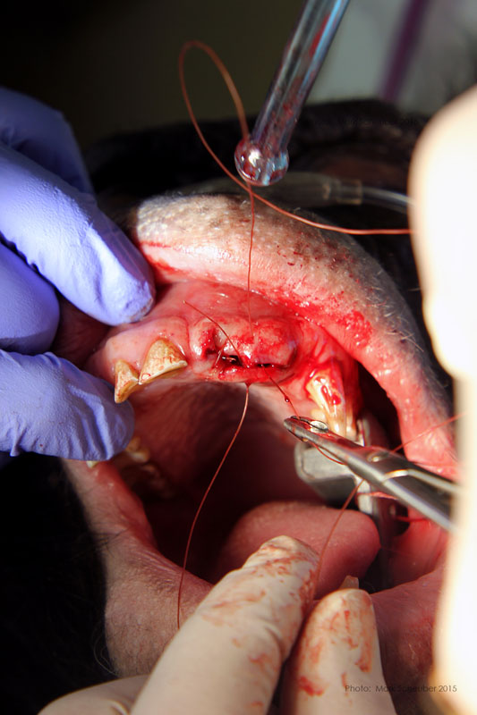
Simple interrupted sutures placed with knots inside the wound so that Makanza will find it difficult to disturb them with his fingers and tongue.
Rachel - Mongolian (Bactrian) Camel
Milwaukee County Zoo.
(Click image to enlarge)
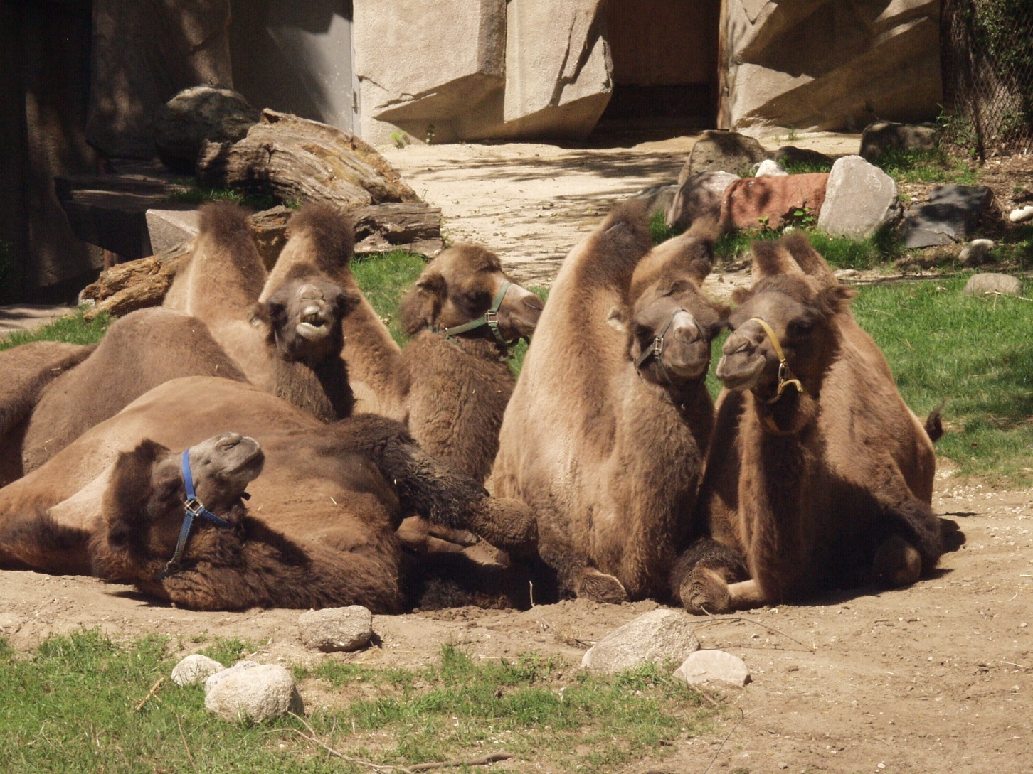
Mongolian (Bactrian) camels
(Click image to enlarge)
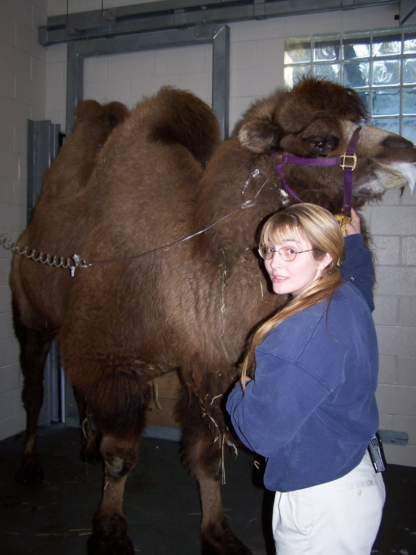
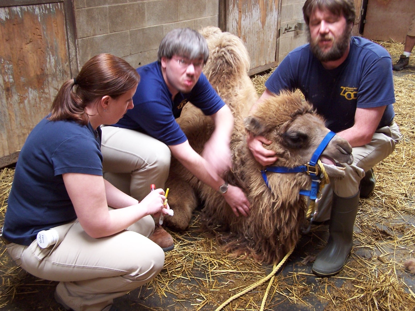
Rachel with keeper Danielle
(Click image to enlarge)
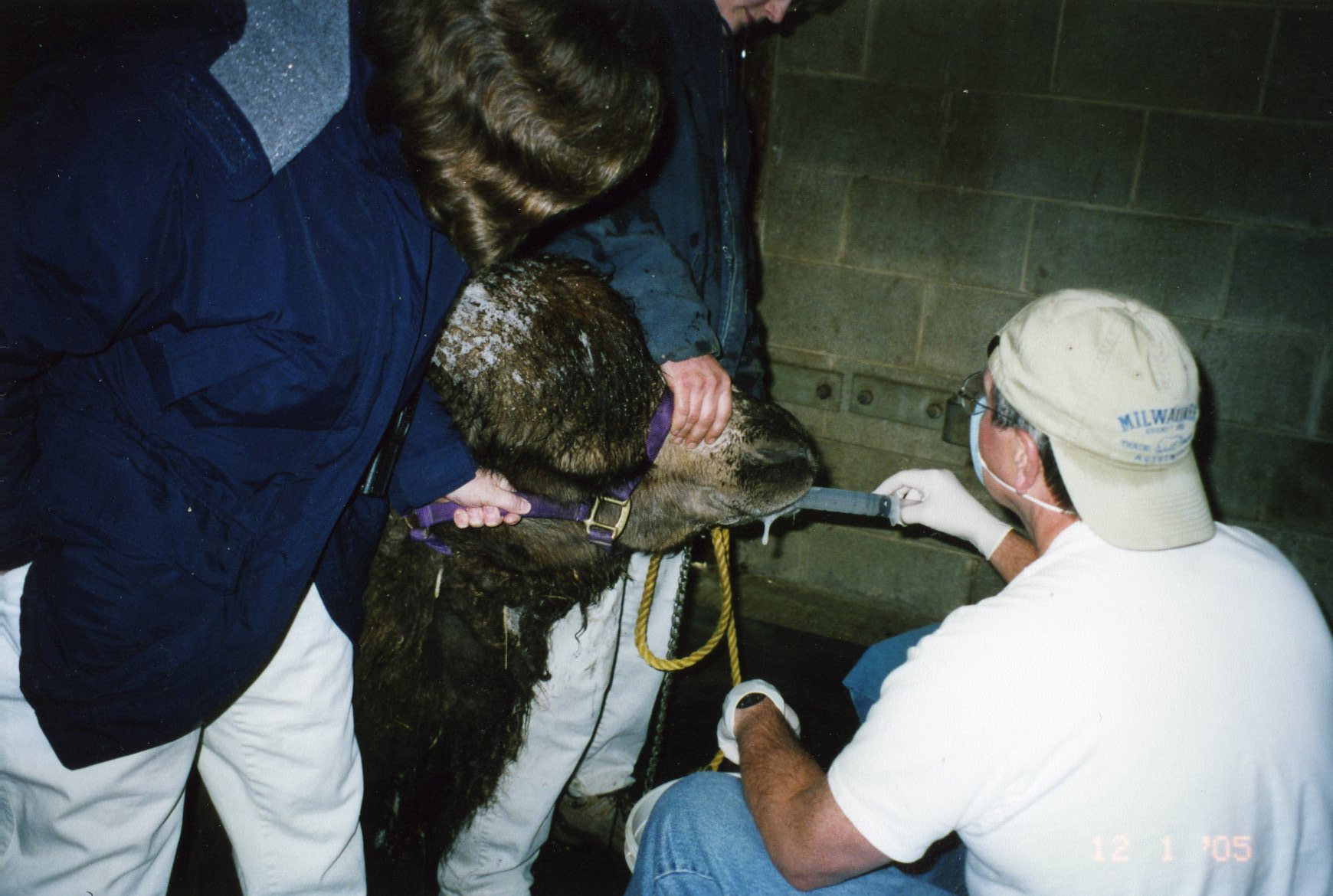
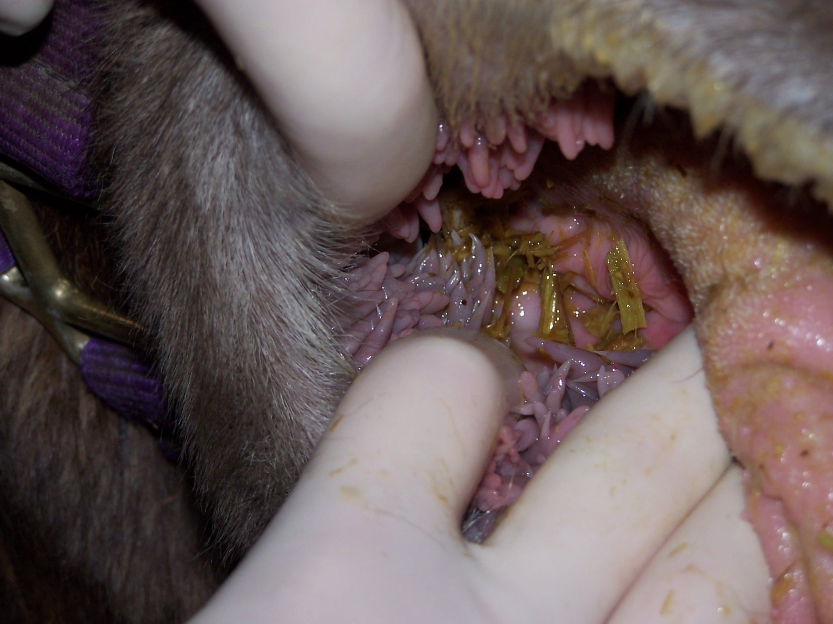
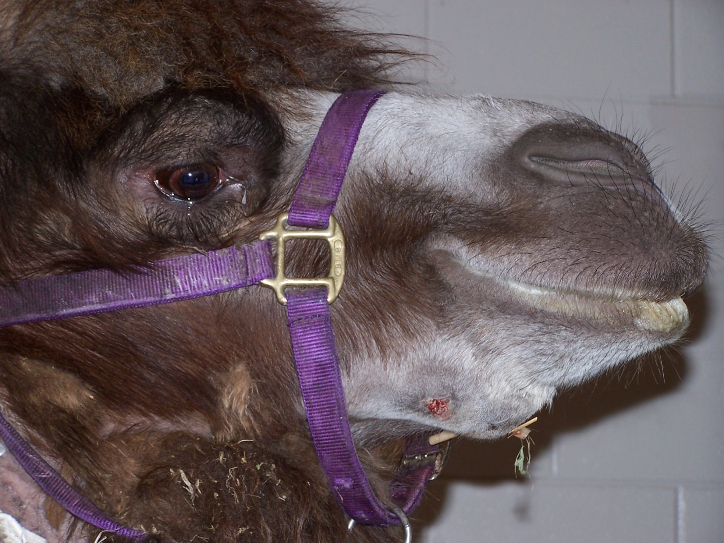
Initial exam, Rachel sedated, flushing out food debris. Note foamy saliva, indicative of oral discomfort, constant tongue action, excessive salivation. Edema over right mandible, premolar area. Promptly identified mobile mandibular right first premolar. Removed easily. Purulent drainage flowed out of the alveolus. At that point, we realized that we had a more serious problem than just one relatively small painful tooth.
(Click image to enlarge)
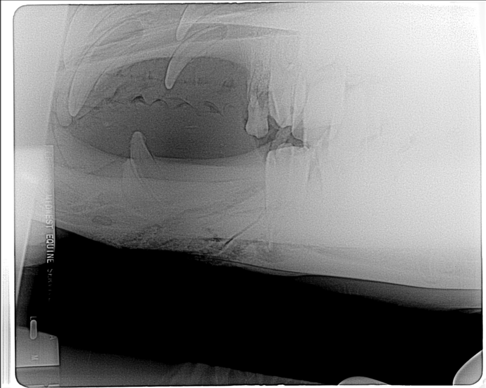
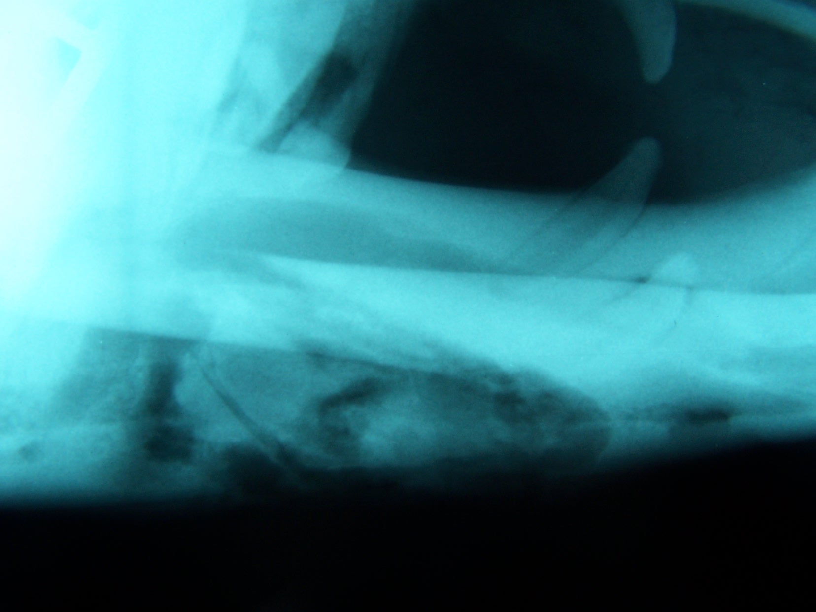
Initial radiograph, taken after I had removed the small first premolar. Purulent exudate flowed out of alveolus. Fracture line along mesial border of second premolar. Antibiotics administered, animal allowed to recover while we discussed further treatment.
(Click image to enlarge)
Radiograph of fractured mandible. Fracture line followed mesial side of second premolar.
(Click image to enlarge)
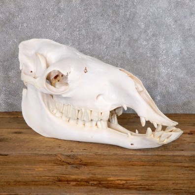
Bactrian Camel Skull.
(Click image to enlarge)
Inraoral view. Note papilla on cheek. Food packed in area of pain by patient, in attempt to relieve discomfort.
(Click image to enlarge)
Abscess pointed, it was explored and drain placed.
(Click image to enlarge)
Radiograph, illustrating progression of osteomyelitis, bone degeneration.
(Click image to enlarge)
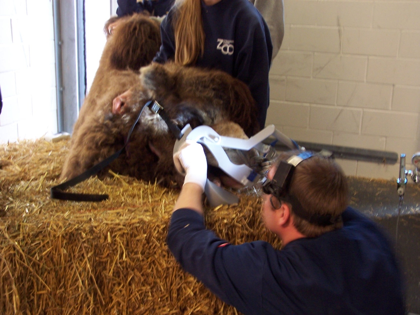
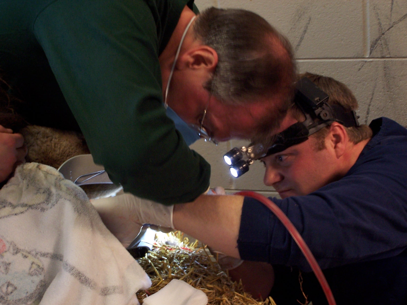
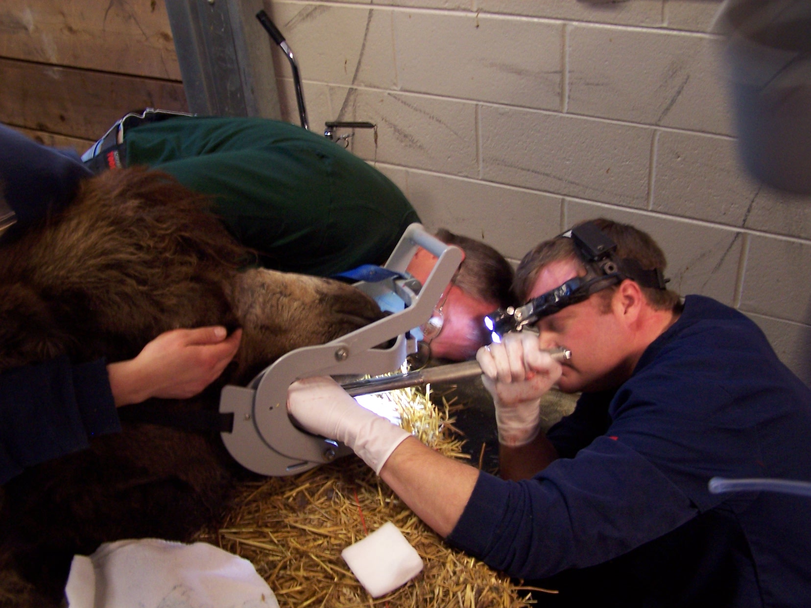
Dr. Travis Henry, a board certified veterinary equine dentist, placing an apparatus to keep mouth open for visualization and treatment.
(Click image to enlarge)
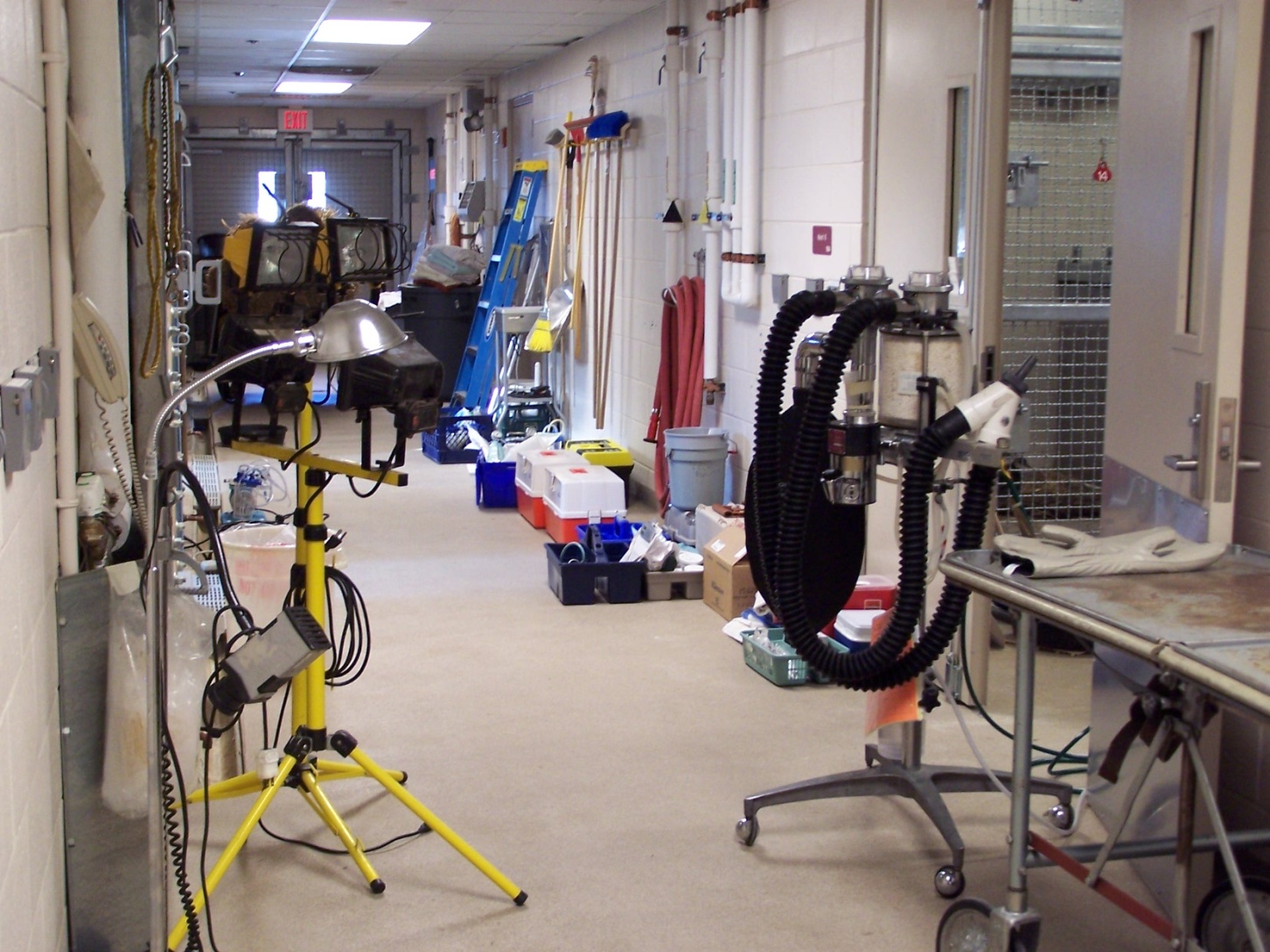
Hall of ICU area, equipment set up.
(Click image to enlarge)
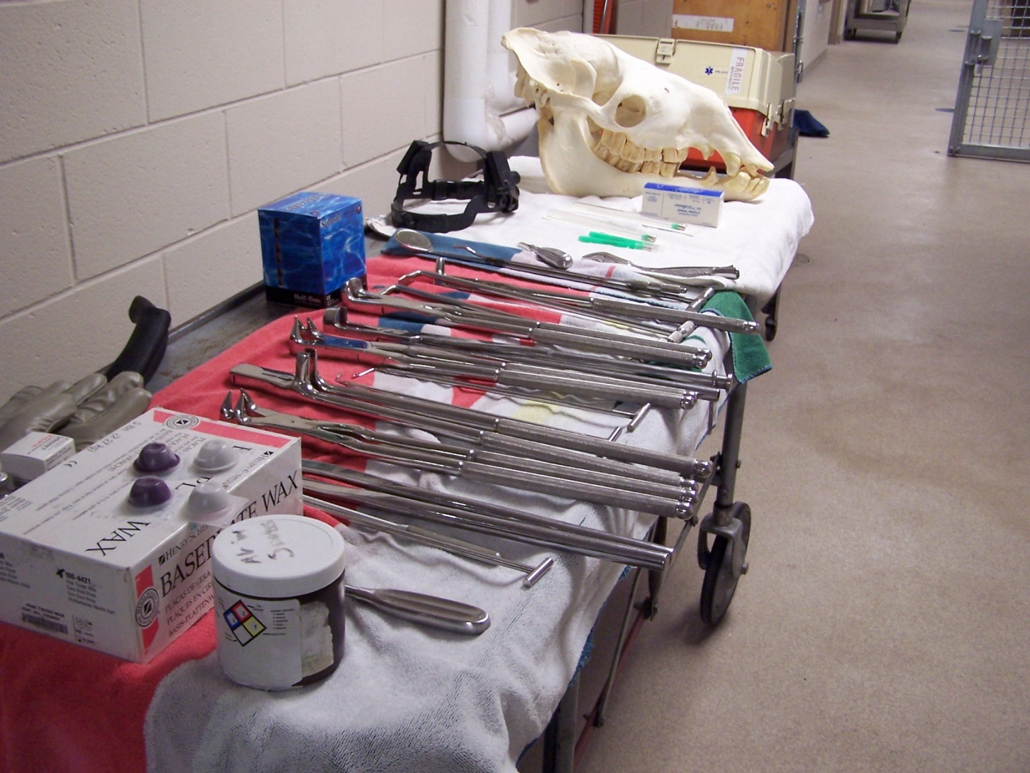
Equine instruments, note Baseplate Wax. Camel skull.
(Click image to enlarge)
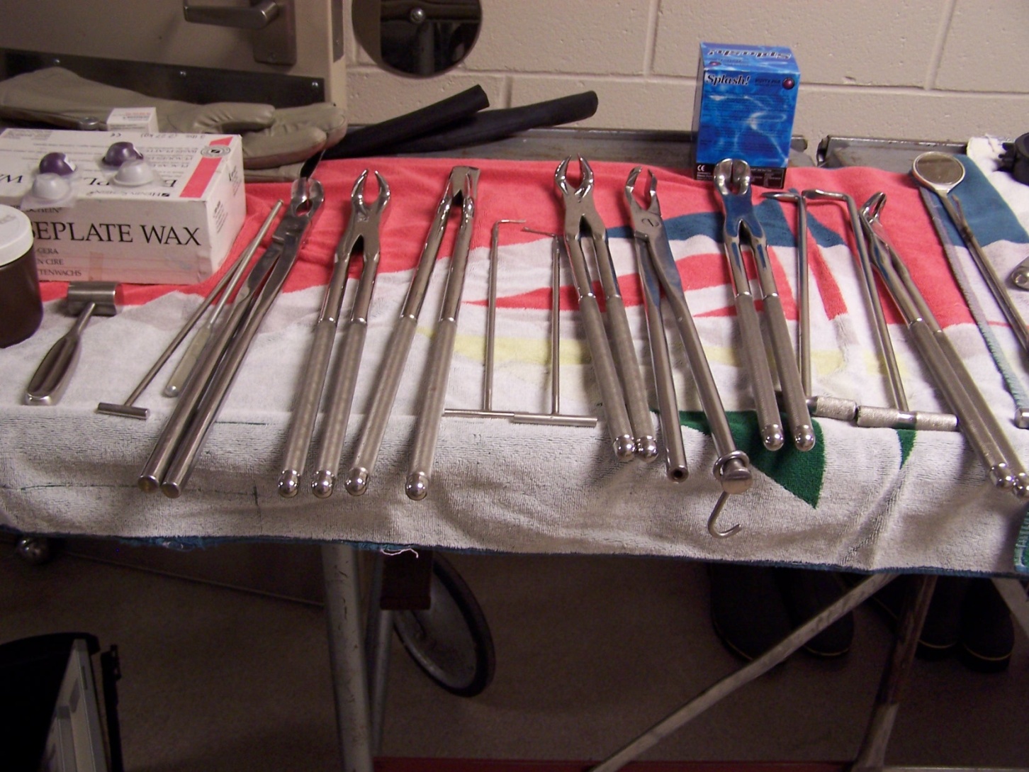
Extraction forceps.
(Click image to enlarge)
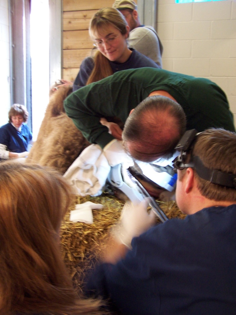
Dr. Henry examining area, Scheels assisting, retracting.
(Click image to enlarge)
Forceps in use.
(Click image to enlarge)
Forceps in use.
(Click image to enlarge)
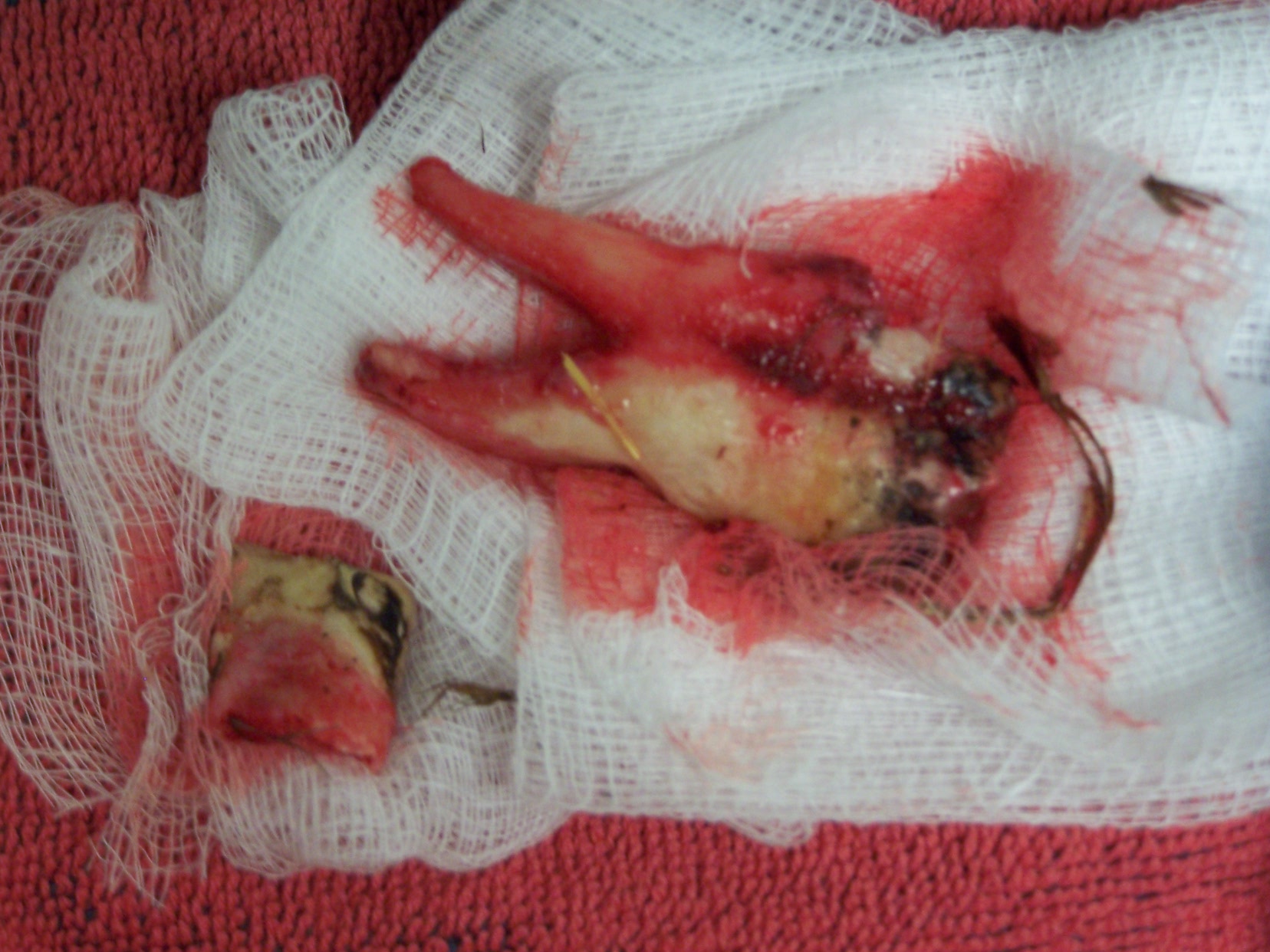
Extracted premolar, fractured crown segment, during removal.
(Click image to enlarge)
Initial radiograph, taken after I had removed the small first premolar. Purulent exudate flowed out of alveolus. Fracture line along mesial border of second premolar. Antibiotics administered, animal allowed to recover while we discussed further treatment.
(Click image to enlarge)
Radiograph of fractured mandible. Fracture line followed mesial side of second premolar. * Find photo of camel dry skull mandible
(Click image to enlarge)
Rachel in Animal Health Center Intensive Care/Quarantine stall.
(Click image to enlarge)
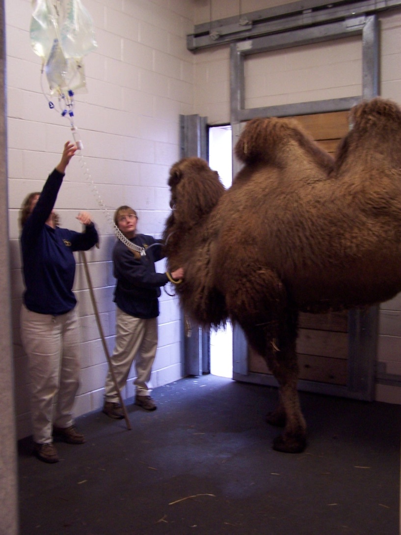
Keepers assisting in sedation procedure.
(Click image to enlarge)
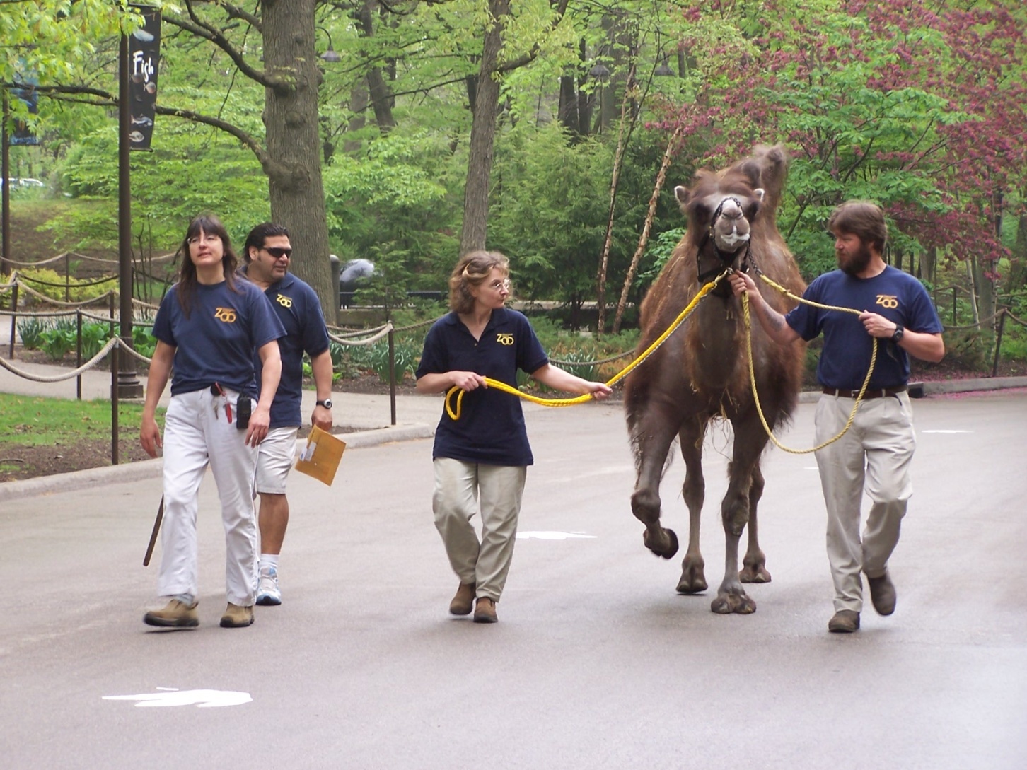
Keepers returning Rachel to the camel area following ? months intensive care. The next day she kicked Robert and fractured his tibia.
This page:
Siberian Tiger Mandibular Canine Endodontic Procedure
Bonobo Makanza
Mongolian (Bactrian) Camel