


Herbivores
Herbivores often exhibit a significant alveolar bone hypertrophy in reaction to jaw bone or tooth pathoses and trauma. The common term "lumpy jaw" has been used to describe the condition. It is a poor term, but we are stuck with it.
Over the years, I believe the most common questions regarding exotic animal dentistry that I have received has been about how to deal with this type of pathosis. I have learned from treating many such cases and from other zoo dental consultants, in particular Drs. Peter Kertesz and David Fagan. I have referenced papers they have either referred to or authored.
This pathosis was described in detail by Sir Frank Colyer in his classic text, Variations and Diseases of the Teeth of Animals, first published in 1936 and updated in 1993. I recommend that anyone serving as an exotic animal dentist acquire this book. I continue to refer to it often.
I had heard the term "lumpy jaw" many times before I encountered an animal that suffered with the problem. If fact, my first experiences with it were the skeletal remains of Dall sheep that had died at Milwaukee County Zoo! (Figure 1)
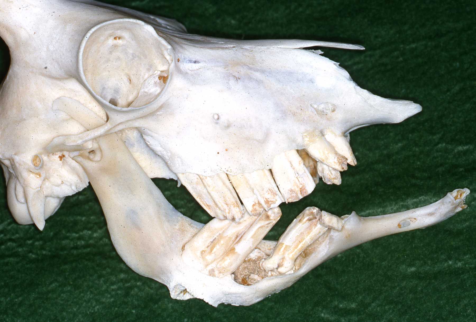 Figure 1
I searched for information regarding this malady. I learned that for many years it had been treated with local antibiotics and relatively superficial treatment of the external wounds associated with the swelling. In many herbivores, such as ruminants, oral antibiotic therapy is ineffective, of no value. In many of these described cases the etiology of the pathosis was not addressed, which I considered quite curious.
In fact, the disease was often referred to by the dominant bacteria, such as Actinomyces bovis in bovine cases. References will still be encountered that refer to the bacteria, although known to be commonly found in the healthy bovine mouth, to be the causative agent of the disorder. Dr. Kertesz pointed out in his 1989 presentation, at the AAZV associated dental conference, that investigators had attributed these complex bacterial infections to exacerbation when an open wound developed and then was exposed to additional pathogens from the environment.
At Milwaukee County Zoo we have learned that a very prompt, aggressive approach is absolutely necessary to optimize a positive outcome in these cases. We periodically hold in-service seminars with our zoo keepers, teaching them what to be looking for while managing their animals. They have been very impressive in observing early signs of pathosis. Early detection increases the potential for a successful treatment outcome. I have listed the signs of note in this sites' introduction. In herbivores, the key sign of bony swelling indicates the animal already has a significant pathosis. Other signs include how the animal is eating, taking a very long time to eat, dropping food, using an unusual head posture to masticate, losing condition or becoming thin. Note, the swelling doesn't ALWAYS occur! Therefore, the other signs must also be considered significant enough to warrant an exam. THAT is the only way to aggressively diagnose and successfully treat these serious pathoses.
I believe that another key element in dealing with herbivores is that these species appear very stoic. I believe they have evolved to attempt to hide any illnesses as part of their "prey" (versus "predator") demeanor. This partly explains why the problem is often quite advanced by the time it is discovered. And remember that we are often dealing with a geriatric population in zoos, so it is not surprising to eventually encounter these types of pathoses manifesting in zoos.
Treating the herbivore dentition presents a few unique treatment challenges. First of all it is simply much more difficult to do a thorough intraoral exam in an herbivore because of their typical small mouth orifice, compared to a primate or carnivore, into a comparatively long oral cavity. Consider how important horses have been to human society for so long and that equine dentistry has finally been developing comprehensive knowledge and treatment approaches just over the last 20 years!
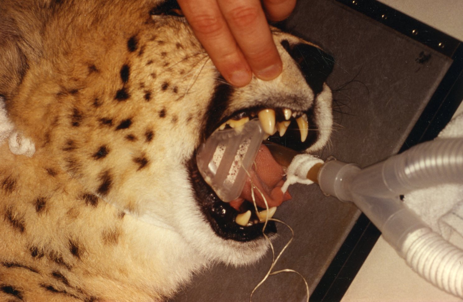
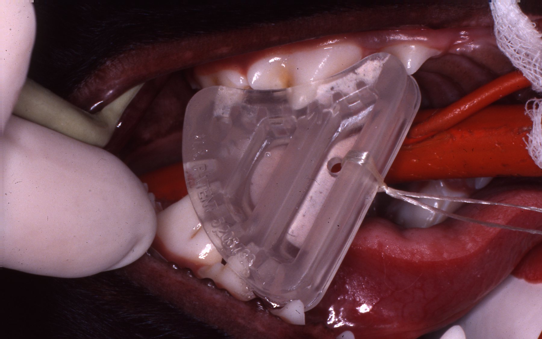
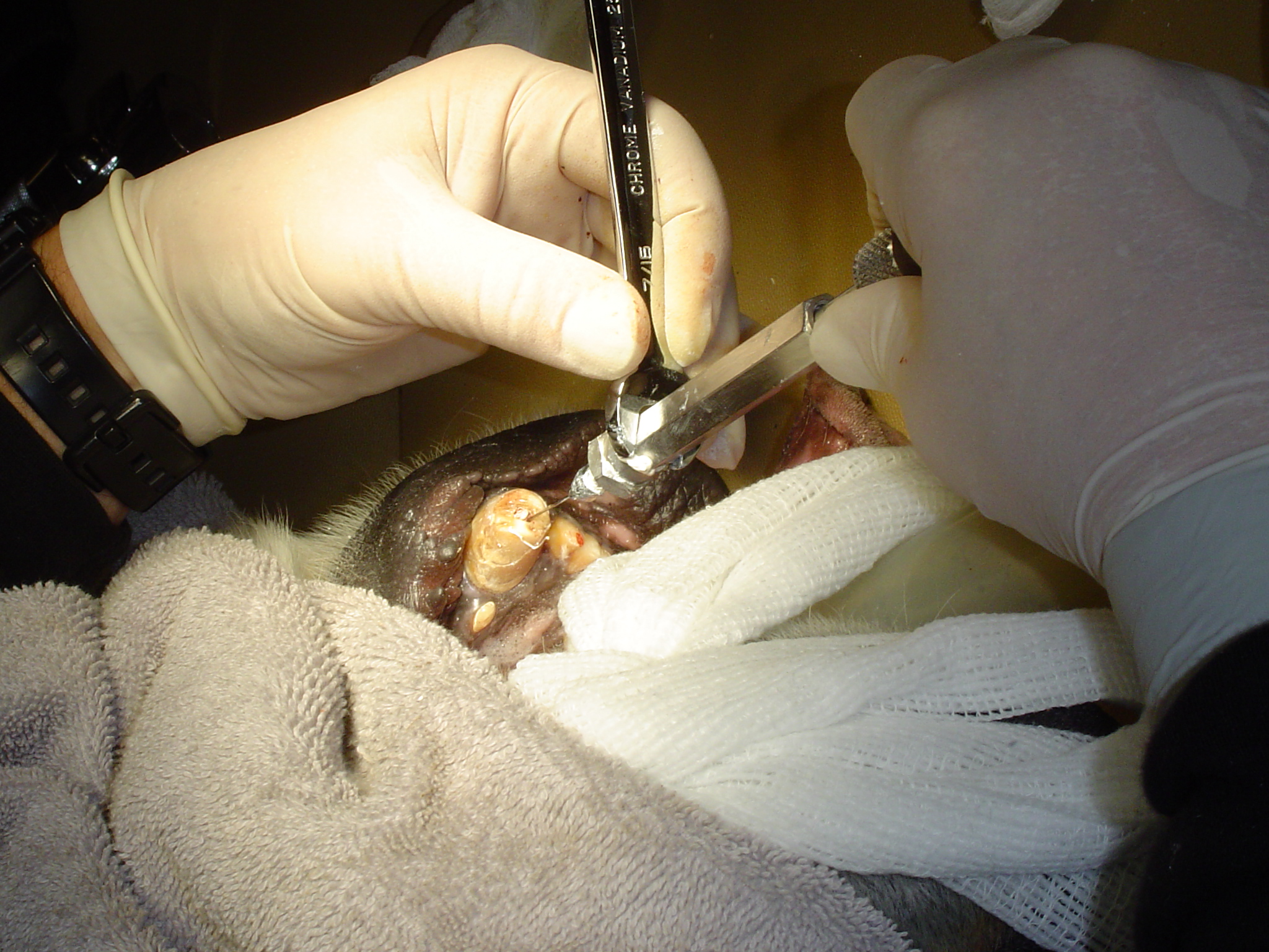
The most common etiology to dental and oral pathosis in herbivores that I have seen is complications related to a fractured tooth. This may lead to pulpal pathosis resulting in abscessation and periapical lesions. A fractured tooth often also results in chronic food impaction, resulting in periodontal pathosis. I have also often encountered periodontal pathosis associated with food impaction due to malocclusion. These are often related to primary tooth retention causing the malocclusion. (Figures 2, 3, 4)
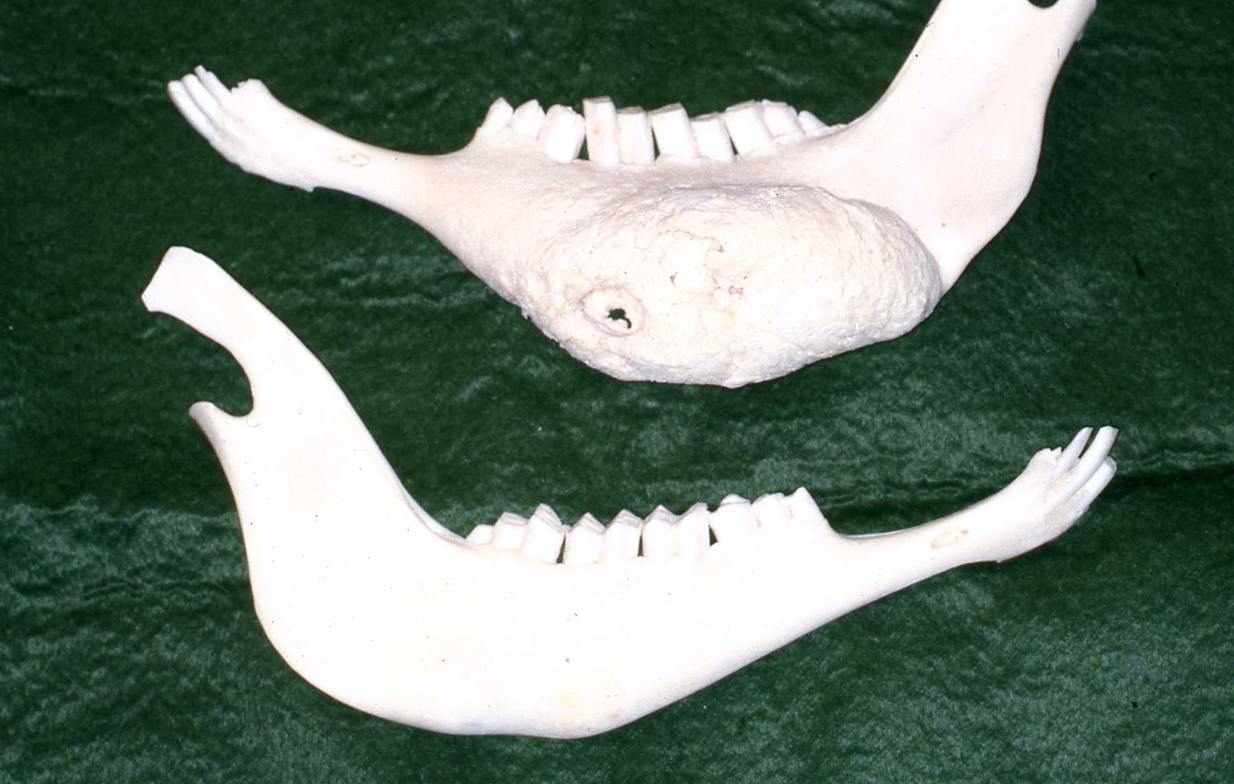 Figure 2
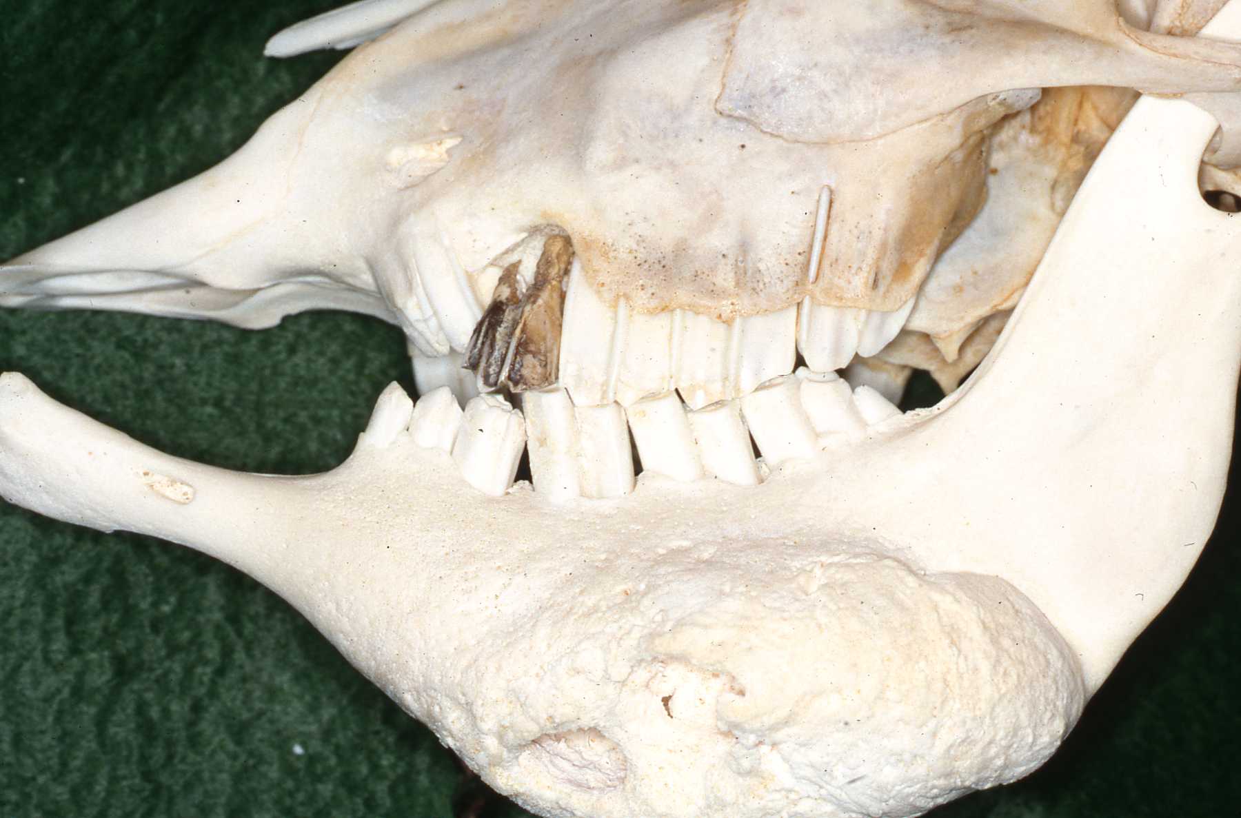 Figure 3
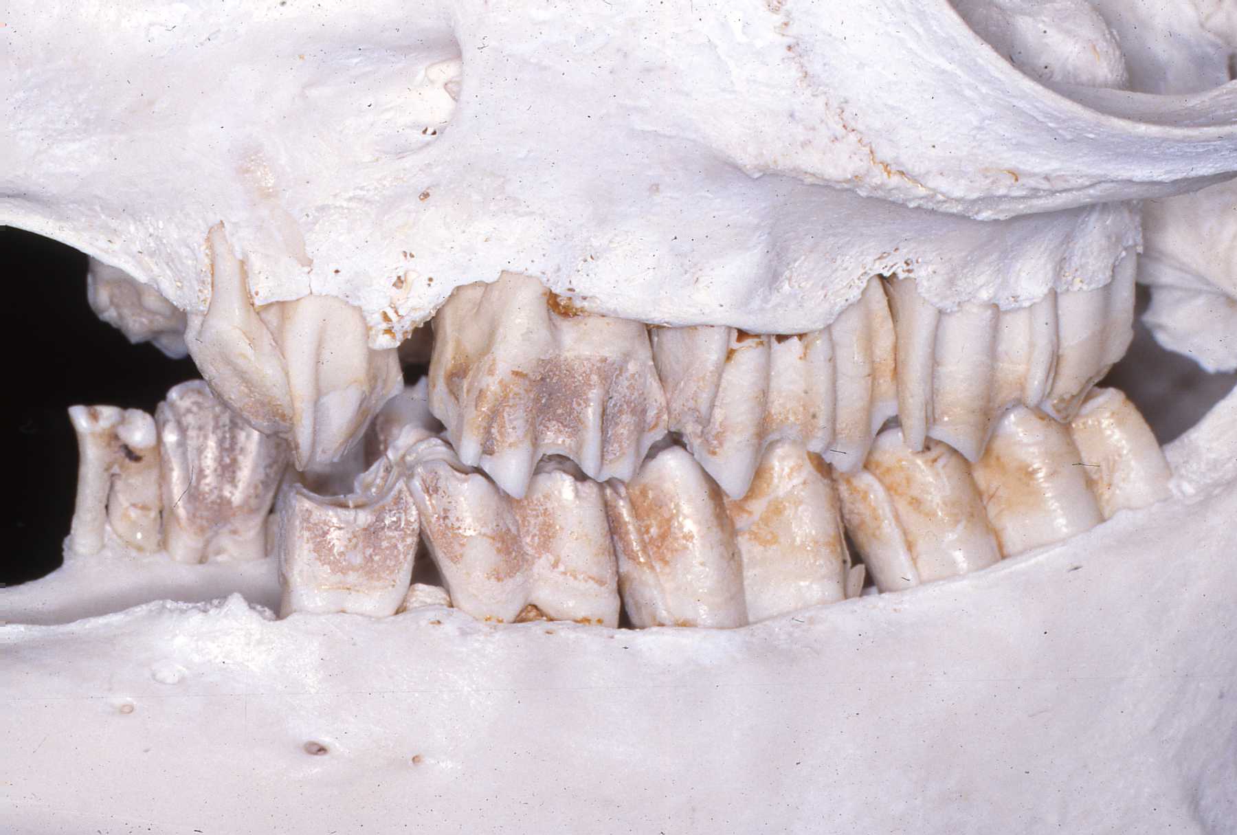 Figure 4
Commonly, food impaction, even in a relatively good dentition, due to "stem" impaction is considered an initiator of the periodontal disease. I agree. This situation is often mentioned in captive animals that may be over crowded, especially macropods. Overcrowding may create food choice or opportunity issues. A natural history example reference regarding Dall sheep refers to the less dominant groups of wild sheep having to forage in less desirable food areas, exhibiting a higher prevalence of oral pathosis. This is attributed to the poorer quality of food choices, being coarser and more inclined to initiate periodontal lesions. Even in these wild groups, the incidence noted is over 25%. And the author told me that he only examined the mandibles! (Hoefs, M. and Bunch, T.D., "Lumpy Jaw in Wild Sheep and its Evolutionary Implications" in Journal of Wildlife Disease, 37(1) 2001. PP 39-48)
The herbivores' posterior teeth, premolars and molars, commonly referred to as "cheek teeth" function as a group. This tooth grouping is often referred to as an arcade due to their intimate positioning, a common shape in a tight row that mimics a common historic European architecture of building rows. They are also categorized as "hypsodont", which means tall teeth. (Figure 5)
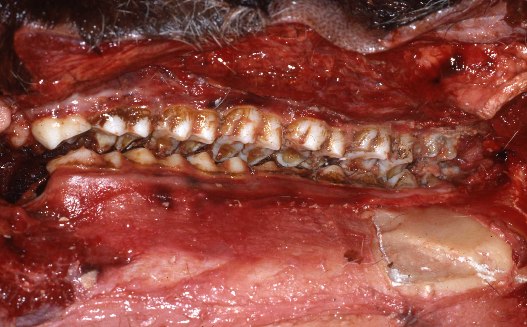 Figure 5
These teeth must thoroughly masticate large volumes of tough fibrous plant foods, chewing them multiple times in ruminants. This is necessary due to the relatively lower food value of plant stems and leaves. The arrangement of enamel, dentin and cementum on the occlusal surfaces in these animals provides a very efficient, grinding surface from first premolar to last molar. Ruminants also have flexible cartilaginous mandibular symphises further contributing to masticatory efficiency.
The equid occlusal surface is also comprised of enamel, dentin and cementum in an even more complex arrangement. Their contrasting hardness coefficients results in a serrated cutting surface of three levels. Another equid tooth specialization that compensates for only one opportunity to masticate is the continual eruption of teeth throughout life. This ongoing eruption provides new tooth structure, maintaining vertical occlusal dimension, as the teeth wear through their life span or until the entire crown is worn away. This is considered to be the optimum efficiency in chewing fibrous food. This most highly evolved situation benefits their hindgut digestion, having only one opportunity to masticate their diet, compared to the ruminants.
Other species presented with the challenge of high volumes of tough fibrous food have evolved teeth arrangements that migrate forward, replacing worn occlusal surfaces with new sharp surfaces. These species include elephants, manatees, and warthogs. This physiological phenomonen in macropods was described by Colyer as "mesial drift".
All of the above mentioned specializations must be thoroughly considered when deciding treatment choices. (Janis, C.M. and Fortelius, M., "On the Means Whereby Mammals Achieve Increased Functional Durability of Their Dentitions with Special Reference to Limiting Factors" in Biology Review, (1988), 63, pp. 197-230)
Among the earliest cases of severe bony swelling in herbivores that I encountered, were a kangaroo that had what the veterinarian and pathologist described as a "sterile abscess." In other words, when we biopsied the swollen bone area, no pathogens were found. However, we did eventually associate the recurring lesion with a malocclusion due to an abnormal permanent tooth eruption situation. We encountered another couple such cases over time and wondered about their etiology.
In 2002 at the AAZV associated dental conference, one of Drs. Fagan and Oosterhuis' presentations attributed such lesions to trauma to tooth buds or immature erupting teeth alveoli. When they pointed this out, it became very clear to us that this was indeed very likely a plausible cause of the lesions that we had been so puzzled about. An example that they mentioned was animals injuring these vulnerable areas when struck against a drinking vessel or food containers' vertical surface. This makes a lot of sense to me when comparing to the natural wild situation of animals drinking from a natural body of water. We prefer to have enclosure drinking and eating stations without vertical surfaces, level with the surface the animal is standing on. This type of husbandry consideration is of significant value in avoiding such injuries. (Fagan, D.A., and Oosterhuis, J.E., "'Lumpy Jaw'"-Another Perspective", The Colyer Institute)
This page:
Discussion
Page 2: Cases